Alzheimer’s Can Begin Changing the Brain Decades Before Memory Loss: New Study Unveils Earlier Indicators
Alzheimer’s disease, a progressive neurodegenerative disorder and the most common cause of dementia worldwide, has long challenged the medical community with its insidious onset and relentless progression. Traditionally, researchers have associated the earliest detectable biological sign of Alzheimer’s with the buildup of amyloid plaques—clusters of misfolded proteins in the brain. Yet, groundbreaking new research suggests the disease’s footprint may begin to alter the brain several years, if not decades, before these plaques can be seen using the most advanced imaging technologies.
This revelation, emerging from a 2026 study published in Nature Neuroscience, prompts a fundamental reappraisal of how, and just as importantly, when Alzheimer’s disease truly begins. The research team from the University of Oslo has identified patterns in brain structure that precede amyloid accumulation by at least seven years—forcing the scientific community to reconsider the earliest indicators of this devastating condition.
The Hidden Early Onset of Alzheimer’s Disease
Alzheimer’s disease is often termed a “silent” disease for good reason. Neuropathological features such as amyloid plaques and tau-based tangles typically accumulate secretly within the brain 15 to 20 years before any obvious memory problems surface. By the time a person seeks medical help for cognitive decline, more than a decade of invisible but destructive brain changes have likely already occurred.
The prevailing research consensus, as highlighted in the recent Nature Neuroscience paper, defines the appearance of significant amyloid-beta plaques as the primary biological marker for an Alzheimer’s diagnosis. This framework has inspired imaging protocols in which PET (positron emission tomography) scans reveal amyloid build-up long before noticeable symptoms manifest. According to this model, structural deterioration of the brain, as measured by MRI (magnetic resonance imaging), appears only after amyloid reaches pathological levels.
However, the University of Oslo’s study challenges these long-held assumptions. By integrating nearly two decades of longitudinal MRI data with sophisticated amyloid PET imaging, the researchers found measurable differences in the cortical thickness—the thickness of the outer layer of the brain—years ahead of amyloid plaque detection. Significantly, these subtle changes were present among older adults who were still cognitively healthy and unaware of the disease brewing within.
Anders M. Fjell, Ph.D., a prominent neuroscientist and co-author of the study, explained that the findings focused on “changes in brain structure in the years before the first scan revealed plaques.” This timeframe extends well beyond the traditional window in which amyloid first appears and into an era when patients feel and function normally in everyday life.
How Brain Scans Reveal Preclinical Changes
PET imaging for amyloid plaques represented a technological leap in Alzheimer’s research, allowing scientists to visualize key pathological structures during life rather than just at autopsy. However, PET scans can only detect plaques once they accumulate above a certain threshold, making them an indicator but not necessarily the true origin point for the disease’s cascade.
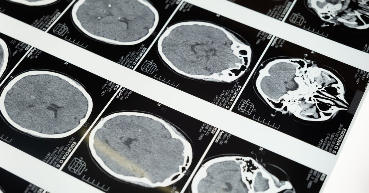
MRI scans, on the other hand, map the structure of the brain, measuring features like cortical thickness and the overall size of critical regions such as the hippocampus, the brain’s memory hub. Until recently, such structural changes were considered to occur after amyloid had begun damaging brain cells—believed to be a sign of disease progression rather than initiation.
Yet, the Oslo team’s analysis suggests this sequence may be inverted. By reviewing thousands of MRI images spanning up to 20 years, the researchers identified individuals whose brains revealed unusual patterns of cortical thickness several years before their PET scans turned positive for amyloid. Remarkably, these pre-amyloid differences diverged from expected aging trajectories and could be seen in participants as much as seven years in advance.
Further complicating the picture, the differences in cortical thickness endured even after accounting for the actual levels of amyloid buildup seen in PET scans. This implies that at least some of these structural alterations may be occurring independently of measurable amyloid, hinting at other potential triggers for the earliest phases of Alzheimer’s pathology.
Unraveling the Causes: Two Competing Theories
The implications of these findings extend far beyond mere early detection. Understanding the drivers of these subtle structural changes could revolutionize how researchers approach Alzheimer’s prevention and therapy.
One theory suggests that amyloid-related processes begin well before they are detectable by current PET technology; the plaques are there, just at concentrations too low to be seen. If correct, this would mean the disease’s initiating events align with existing models, and technological advances may simply allow us to recognize them earlier.
The more disruptive hypothesis is that a different molecular or cellular process initiates brain changes before amyloid takes the central role. If so, this would indicate that focusing only on amyloid in both research and drug development could mean missing a window for even earlier, potentially more effective interventions.
Dr. Fjell underscores this uncertainty, noting, “If the latter is true, it suggests it’s important to continue developing drugs that target processes other than amyloid plaque accumulation.” This reflects a growing call in the field for a broader investigation of disease mechanisms, encompassing inflammation, vascular contributions, tau pathology, and genetic risk, among others.
International and Scientific Context: Expanding Evidence for a Longer Preclinical Phase
While the Oslo study stands out for its rigorous methodology and unusually long timeline, it is not alone in suggesting Alzheimer’s disease incubates for years before overt symptoms or detectable plaques. Several international studies have confirmed that pathological changes in the brain often precede clinical diagnosis by a decade or more.
A notable genetic study in Alzheimer’s & Dementia observed that individuals at high risk for familial Alzheimer’s experienced elevated amyloid-beta levels up to 22 years before symptoms emerged. Similarly, a large prospective study published in Neurology, tracking over 2,000 Chicago residents, found cognitive shifts in memory and executive function as early as 18 years before an official dementia diagnosis.
Taken together, these studies underscore the importance of monitoring for the earliest, most subtle changes—whether in cognitive performance or brain structure—in the quest to develop interventions capable of halting or slowing the disease before substantial damage occurs.
Why This Research is Especially Significant
Most studies of Alzheimer’s rely on relatively short follow-up periods. Since the disease’s pathogenesis may stretch over 20 years or more, findings from three-to-five-year studies can easily miss the true arc of preclinical changes. The Oslo research’s strength lies in its extraordinary dataset: 4,570 longitudinal MRI scans and 1,684 amyloid PET scans, gathered across three cohorts of cognitively healthy adults, some followed for almost two decades.
This approach allowed investigators to trace backward through participants’ brain imaging records, identifying when structural changes might first set in—well before these individuals crossed the “amyloid-positive” threshold by current diagnostic standards. Lead researcher James Michael Roe, now International Scientific Lead with Cercare Medical, highlighted that “structural changes in the brain occur many years before high levels of plaque are seen on PET scans,” shifting the field’s understanding of what counts as the earliest phase of Alzheimer’s.
Such longitudinal research is rare due to the expense and complexity of maintaining healthy aging cohorts over many years. Its findings add robust evidence to a paradigm shift in how scientists conceptualize Alzheimer’s trajectory from health to disease.
Caveats and What This Study Doesn’t Claim
Though compelling, these findings invite cautious interpretation. The Oslo study, peer-reviewed and published in a leading neuroscience journal, is observational and not yet a basis for clinical protocols or screening recommendations.
Structural MRI changes are common with normal aging and can overlap with patterns seen in other neurological disorders. More extensive validation in larger and more diverse populations is required to confirm whether these patterns are specific to Alzheimer’s risk or merely incidental. Existing diagnostic tools—including a combination of amyloid, tau, and FDG PET, plus detailed neurocognitive testing—will remain the gold standard for now, with the FDA in the United States and health authorities worldwide approving new therapies and imaging techniques based on this framework.
The Oslo team acknowledges that further research is needed to explore how these early changes relate to established risk factors such as genetics, vascular health, inflammation, and the accumulation of tau proteins. More importantly, there is not yet a routine or recommended clinical test for detecting these subtle structural changes in healthy adults, nor is there proof that their presence inevitably leads to Alzheimer’s symptoms.
Implications for Alzheimer’s Research and Prevention
The study’s most important takeaway may not be the theoretical possibility of diagnosing Alzheimer’s years earlier, but rather the urgent need for a re-evaluation of when and how the disease starts. Detecting the disease’s signals sooner could have significant consequences for both drug development and preventive medicine, allowing novel therapies to be deployed before irreversible damage takes place.
Alzheimer’s research and treatment are at a crossroads. As new medications that target amyloid arrive on the market—some of which have received regulatory approval for use in the early stages of disease—scientists must develop clearer definitions for what counts as “early.” If processes beyond amyloid accumulation are altering the brain even sooner, multidisciplinary approaches to treatment and prevention may deliver better results in the future.
For individuals concerned about memory loss or cognitive change, the message remains consistent: consult healthcare professionals and rely on established screening and diagnostic tools. For researchers, the findings emphasize the importance of comprehensive longitudinal studies and advanced imaging to chart the elusive preclinical phase of Alzheimer’s disease.
Disclaimer: This information is intended solely for educational and informational purposes. It should not be used as a substitute for professional medical advice, diagnosis, or treatment. Readers should always consult qualified healthcare providers with questions regarding any medical condition or changes in health.
Sources
- Nature Neuroscience – Cortical thickness changes precede high levels of amyloid by at least 7 years
- ScienceDaily – New imaging study shows Alzheimer’s may begin earlier than previously thought
- Alzheimer’s & Dementia – Amyloid-beta accumulation in autosomal dominant Alzheimer’s
- Neurology – Cognitive decline in older persons in the years before and after incident Alzheimer’s disease
- CDC – Alzheimer’s Disease and Healthy Aging
- WHO – Dementia Fact Sheet
- Mayo Clinic – Alzheimer’s Disease: Symptoms & Causes
- BBC – Major advances in Alzheimer’s detection and treatment
- Reuters – Healthcare & Pharmaceuticals: Alzheimer’s Research
- The Guardian – Alzheimer’s Disease News
Disclaimer: This content is intended for entertainment purposes only and is not based on real events.