
New Bioactive Material Regenerates Knee Cartilage: Breakthrough in Joint Repair
The human body possesses remarkable self-healing powers. However, some tissues, including joint cartilage, exhibit very limited regenerative capacity. Cartilage, especially in the knee, is crucial for painless movement and mobility, yet its poor blood supply makes natural repair challenging. Damage to knee cartilage can lead to chronic pain, restricted movement, and eventually diseases like osteoarthritis. Conventional treatments, ranging from surgical stimulation to joint replacement, often involve lengthy recovery times and mixed results.
Recently, researchers from Northwestern University unveiled an innovative solution: a novel bioactive material that enabled significant regeneration of high-quality cartilage in the knees of animal models. This discovery marks a critical step forward in regenerative medicine and holds potential to transform the management of joint injuries and degenerative conditions.
The Medical Possibilities of Regenerating Cartilage

Cartilage is the shock-absorbing tissue that lines the ends of bones within joints, cushioning movement and ensuring smooth motion. As Samuel I. Stupp, senior author of the study, explains in an interview with Northwestern Now: “Cartilage is a critical component in our joints. When cartilage becomes damaged or breaks down over time, it can have a great impact on people’s overall health and mobility. The problem is that, in adult humans, cartilage does not have an inherent ability to heal. Our new therapy can induce repair in a tissue that does not naturally regenerate. We think our treatment could help address a serious, unmet clinical need.”
In the 2024 animal study, the new biomaterial was applied to cartilage defects within the knee joints of sheep. Astonishingly, within just six months, the treated joints displayed marked cartilage regeneration, with newly-formed tissue closely resembling natural cartilage in both structure and molecular composition. These early results suggest that the approach could, with further development, offer effective solutions for common joint injuries—such as anterior cruciate ligament (ACL) tears—while also potentially reducing the need for knee replacement surgery and improving outcomes for individuals with osteoarthritis.
Osteoarthritis affects hundreds of millions globally, and knee injuries are among the most prevalent musculoskeletal complaints, especially among athletes and older adults. Improved methods for restoring healthy cartilage could vastly improve patients’ quality of life while reducing the enormous healthcare burden associated with chronic joint conditions.
What Is in the Biomaterial?
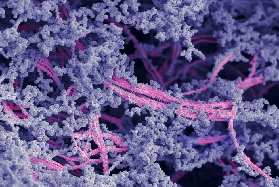
This recent breakthrough builds on years of research by the Stupp Laboratory into bioactive materials. Prior work, including a 2023 study, demonstrated how molecular “dancing molecules” could enhance the production of proteins of cartilage-forming cells. The latest study, however, introduced a hybrid biomaterial composed of two essential components: a bioactive peptide and modified hyaluronic acid.
The bioactive peptide is designed to bind strongly to a critical protein involved in the induction of cartilage growth—essentially acting as a targeted biochemical signal to direct the body’s own stem cells to repair the damaged area. Modified hyaluronic acid, meanwhile, is a substance naturally present throughout the human body. It is best known to the public as a moisturizing ingredient in skincare products, but it also supports and lubricates cartilage and other connective tissues, contributing to joint hydration and resilience.
As described by Dr. Stupp, “Many people are familiar with hyaluronic acid because it’s a popular ingredient in skincare products… It’s also naturally found in many tissues throughout the human body, including the joints and brain. We chose it because it resembles the natural polymers found in cartilage.”
Once mixed, these two ingredients spontaneously assemble into fibrous bundles—mimicking the natural extracellular matrix of cartilage. When introduced into a joint injury, these fibers provide a supportive platform for endogenous cells, sending molecular cues to initiate, direct, and sustain natural cartilage regeneration. Importantly, the goal is not to introduce foreign cells, but to harness and stimulate the body’s innate capacity for repair.
The Test to Regenerate Cartilage: Animal Model Research
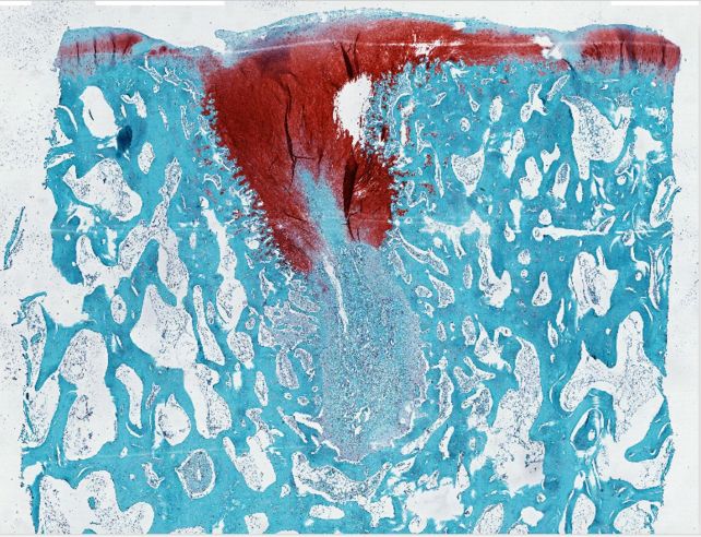
To rigorously evaluate the biomaterial’s effectiveness, the Northwestern research team selected sheep as their animal model. The stifle joint in sheep—the equivalent of the human knee—shares many anatomical and mechanical properties with human joints, including weight-bearing and movement. Crucially, sheep cartilage is notoriously difficult to regenerate, making them an ideal model for challenging joint repair therapies.
During the experiment, researchers surgically induced cartilage defects within the sheep’s stifle joint. The biomaterial was then injected directly into the site of injury, where it quickly transformed into a rubber-like matrix. Over the next several months, as the matrix gradually degraded, it was replaced by new cartilage tissue. Laboratory tests and microscopic analysis later confirmed that the regenerated tissue closely resembled healthy, natural cartilage, not the fibrocartilage or scar tissue that commonly forms in similar conditions.
Dr. Stupp highlighted why this model is particularly predictive for future human applications: “A study on a sheep model is more predictive of how the treatment will work in humans. In other smaller animals, cartilage regeneration occurs much more readily.” The success of this method in sheep provides stronger evidence for its possible effectiveness in human patients, and sets the stage for further preclinical and clinical trials.
Beyond structural observations, biomechanical testing revealed that the repaired tissue possessed similar functional properties—such as elasticity, resilience, and durability—to normal joint cartilage. This is a crucial finding: high-quality regeneration means not only pain relief, but the potential for restored mobility and long-lasting results.
The Future of Knee Treatment and Regenerative Medicine

Looking ahead, the Northwestern team is optimistic that the new biomaterial could be implemented during orthopedic surgeries, potentially revolutionizing current clinical approaches to cartilage injury and degeneration. For many decades, the standard of care has included bone microfracture, a surgical technique in which small holes are made in the underlying bone to encourage the formation of new cartilage. Yet, as explained by MedlinePlus and the Mayo Clinic, this typically results in the formation of fibrocartilage—a structurally weaker form of tissue than the original hyaline cartilage required for normal joint function.
When surgical repair does not restore sufficient function, partial or total knee replacement using artificial implants is often prescribed. While these procedures can alleviate severe pain and restore movement, they carry significant risks, including infection, implant wear, and a lengthy rehabilitation period. Furthermore, they may not be suitable for younger, more active patients who are likely to require several replacements over their lifetime.
“The main issue with the microfracture approach is that it often results in the formation of fibrocartilage — the same cartilage in our ears — as opposed to hyaline cartilage, which is the one we need to have functional joints,” Dr. Stupp clarified. “By regenerating hyaline cartilage, our approach should be more resistant to wear and tear, fixing the problem of poor mobility and joint pain for the long term while also avoiding the need for joint reconstruction with large pieces of hardware.”
If further research—including human clinical trials—proves safe and successful, the implications could be transformative. Patients may benefit from faster recoveries, greater pain relief, improved joint function, and less reliance on invasive surgeries. People living with osteoarthritis, who often face limited options beyond symptomatic treatment, may finally have access to therapies that can genuinely restore joint integrity and mobility.
Experts in orthopedic surgery, regenerative medicine, and tissue engineering have welcomed these findings. Although more research is necessary before widespread clinical adoption, this bioactive material represents a promising new direction in the treatment of joint injuries, with potential applications not only in knees but other load-bearing joints affected by trauma, aging, or disease.
The societal significance cannot be overstated. As the population ages and rates of obesity and sports injuries continue to rise, the need for durable, non-invasive joint restoration methods will only increase. Regenerative therapies such as this one could drastically reduce the economic and quality-of-life burdens associated with musculoskeletal disorders worldwide.
Continued Research and the Road Ahead
While the initial results of this bioactive material are highly promising, several steps remain before it can be introduced into clinical practice. Rigorous safety testing, detailed evaluation in larger and more diverse animal populations, and eventually human clinical trials will be necessary to determine its long-term effectiveness and any potential side effects. Close collaboration between biomedical engineers, clinicians, and regulatory bodies will be crucial to ensure that future therapies meet the highest standards of safety and efficacy.
Simultaneously, scientists are eager to explore whether similar bioactive materials can be fine-tuned for use in other joints, such as hips, shoulders, or even intervertebral discs. There is also active research into combining such approaches with biological factors like stem cells or growth factors, potentially enhancing repair capacity even further.
Public interest in cartilage regeneration and joint preservation therapies is high, as evidenced by increasing investment and global collaborations in tissue engineering. Patient advocacy groups remain hopeful that these advancements will translate swiftly and safely into real-world solutions. Meanwhile, the current research offers hope for millions struggling with chronic knee pain and mobility impairment.
Conclusion
The development of a bioactive material capable of regrowing functional knee cartilage represents a significant milestone in regenerative medicine. Through careful scientific innovation, researchers at Northwestern University have demonstrated that even tissues long thought to be beyond natural repair may one day be restored through targeted biochemical interventions. As the field progresses, such approaches may offer new hope to those suffering from joint injury, degeneration, and pain, paving the way for a future of healthier, more resilient joints.
Sources
- Northwestern Now
- Proceedings of the National Academy of Sciences (PNAS)
- Journal of the American Chemical Society (JACS)
- MedlinePlus
- Mayo Clinic
Disclaimer: This content is intended for entertainment purposes only and is not based on real events.